October 2019
INTERESTING ECHO OF THE MONTH
Case History
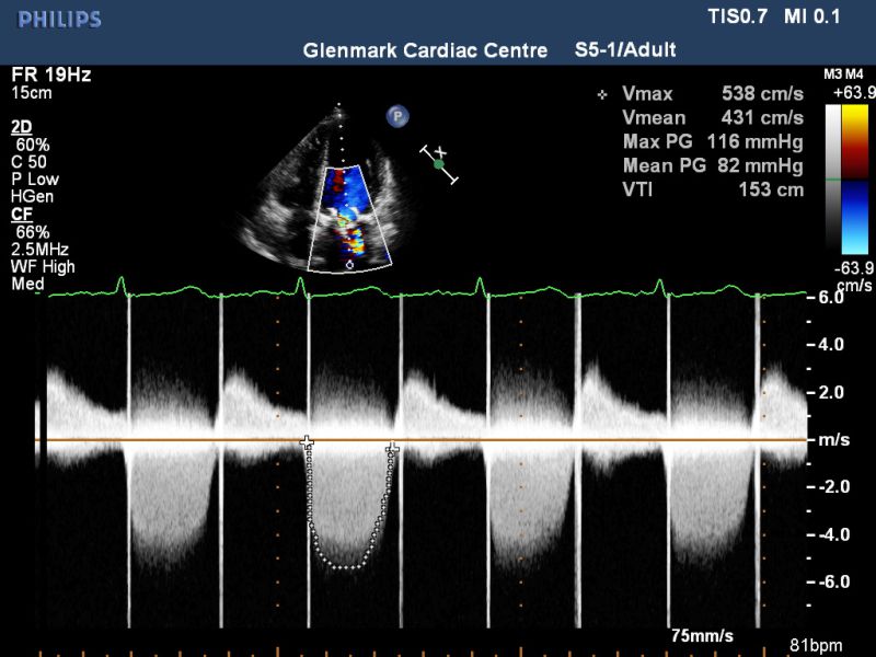
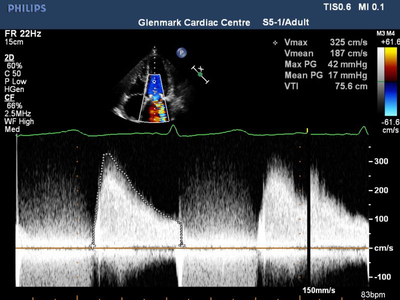
A 65 years male with a history of MVR in 2009 with a Bio prosthetic Mitral Valve, presented with recent onset of DOE NYHA class III (No history of Fever). His echo clips are shown. What is the Diagnosis??
Degenerated Bioprosthetic mitral valve dysfunction with flail AML.
The echo clip of apical four chamber shows thickened and calcific mitral valve leaflets with evidence of a flail anterior leaflet which prolapses into the LA in systole. The colour Doppler four chamber view shows a significant intravalvar mitral regurgitation and the CW Doppler confirms the MR and also shows a high gradient across the prosthetic mitral valve ( peak / mean = 42/17 mm hg).
INTERESTING ECG OF THE MONTH
Case History
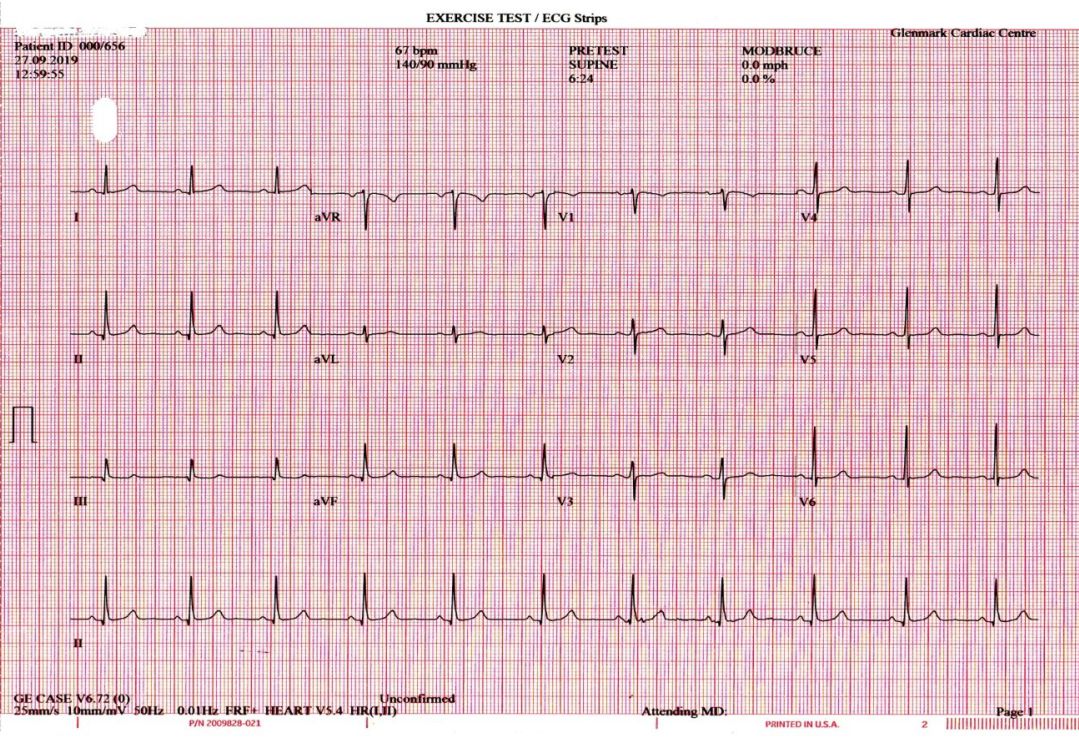
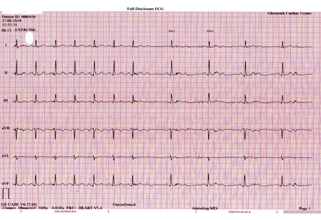
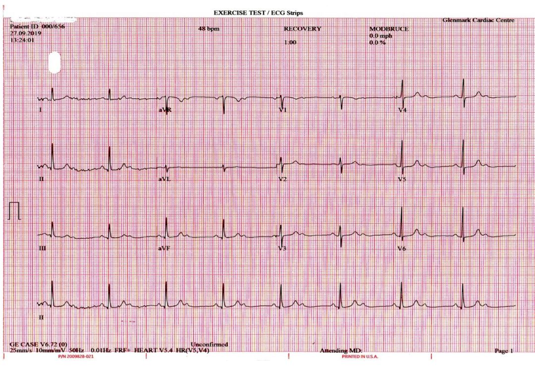
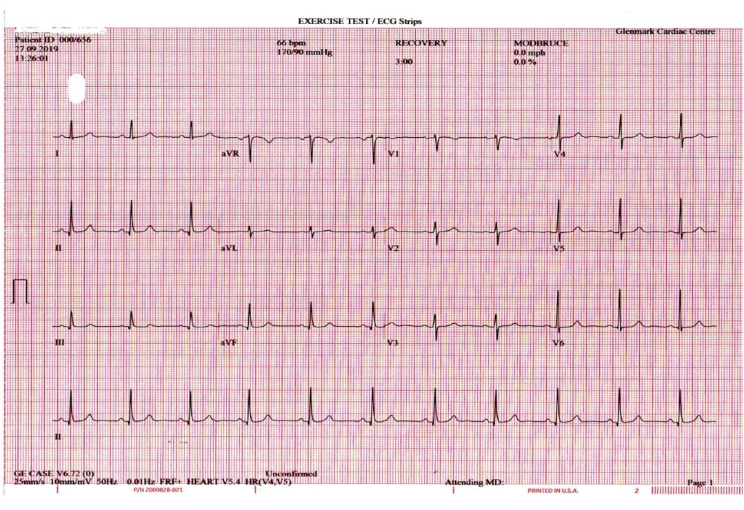
A 65 yrs female presented with dyspnea, NYHA II . She was advised treadmill stress test to rule out coronary artery disease. Above are her ECG tracings during the stress test. What do the ECGs show? Explain the mechanism of the abnormality seen.
The basal ECG shows SR with a rate 67 bpm and narrow QRS. At peak exercise the patient developed breathlessness, when the heart rate was 85 bpm. Subsequently there was a sudden slowing of the heart rate to 40 bpm due to 2:1 AV block. The QRS was narrow during the AV block, which lasted till 2 minutes of recovery. 1:1 AV conduction resumed when the sinus rate reduced to 70 bpm. The level of AV block is likely below the AV node as there is worsening of AV conduction with increased sympathetic tone & sinus rate during exercise and improvement of AV conduction at slower sinus rate during recovery. Because the QRS is narrow throughout, the AV block is most likely within the His bundle. The etiology of the infra Hisian block could be degenerative or due to coronary artery disease with ischemia of the artery supplying the His bundle (more often from the LAD artery).

