June 2025
INTERESTING ECHO OF THE MONTH
Case History
34 yrs-old male, S/P AVR (St Jude mechanical valve) in 2020. Presented with dyspnea on excretion . Increased symptoms since one month. Had a parieto-temporal stroke on sept, 2023. No history of fever.
Physical findings: BP = 90/60 mm Hg, pulse = 96 beats /min, O2 = 98 %, JVP not raised, no pallor/thyroid. CVS – ESM +EDM, Chest – clear.
ECG: LVH with strain.
What is the Diagnosis??
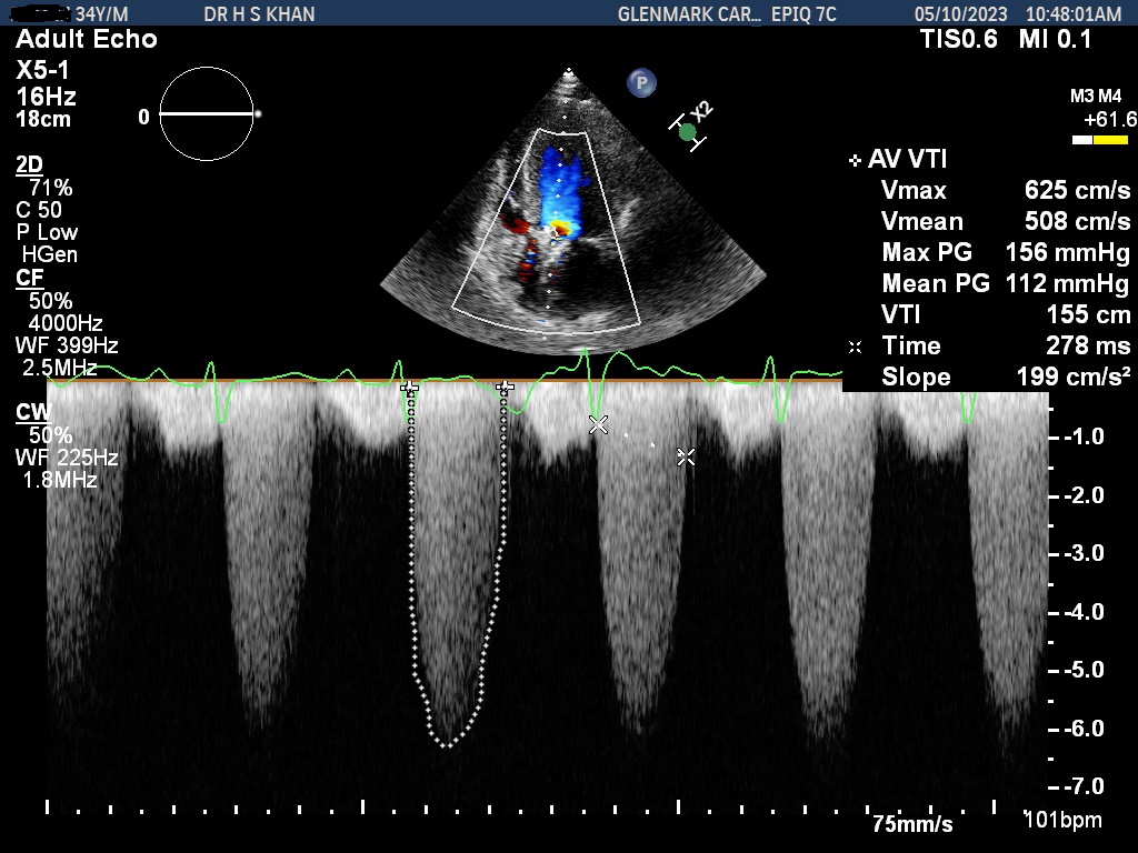
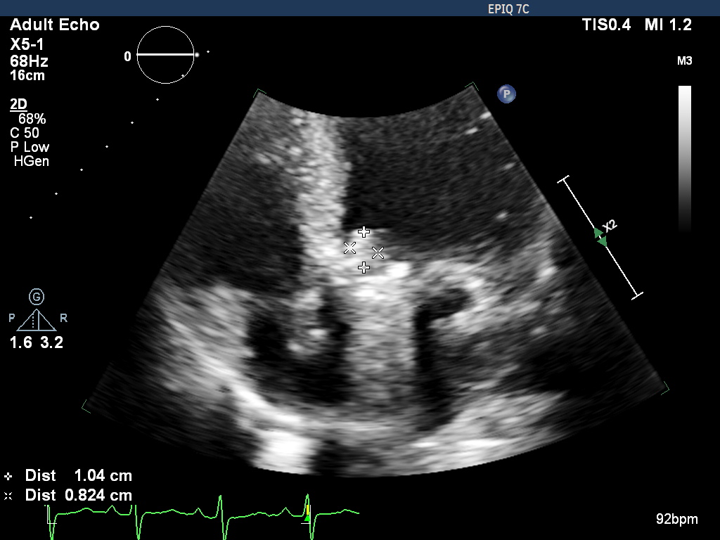
Impression: Metallic aortic valve thrombosis in a recent CVA
The Echo clips in apical 5 chamber view show there is a metallic prosthesis in aortic position. There is severe aortic restenosis. The peak / mean systolic gradient across the aortic valve are 156/112 mm Hg. The aortic valve area by continuity is 0.7 sq cm. There is moderately severe (Grade III/IV) intravalvar leak. There is a suggestion of thrombus on aortic valve measuring 12 x 18 mm. Echo features are suggestive of a stuck valve