August 2019
INTERESTING CASE OF THE MONTH


Case History
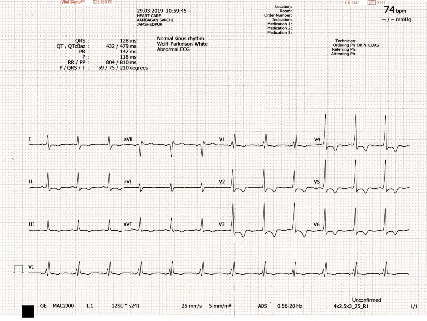
A 55 year old male patient presented with angina and dyspnea on exertion, NYHA class III, gradually worsening over 6 months . His ECG revealed RBBB and deep, symmetrical T wave inversions in precordial leads. His echo clips are shown. What is the diagnosis??
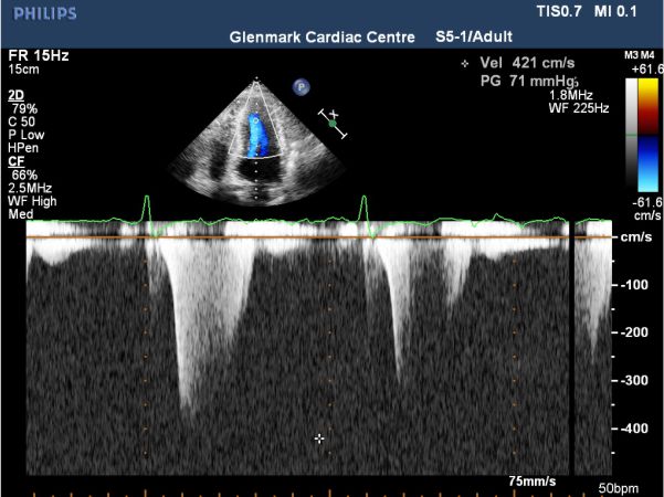
Apical hypertrophic cardiomyopathy. The echo clip of the AP 4 Chamber view shows significant hypertrophy of left ventricular apical segments with near obliteration of LV cavity, during systole. There is a flow acceleration seen in the apical LV cavity with an intracavitatory gradient of 71 mm Hg recorded. There is no SAM of the mitral valve or dynamic LVOT gradient in the PLAX view. The SAX view confirms the apical cavity obliteration and highlights the significant apical LV hypertrophy.
