February 2023
INTERESTING ECHO OF THE MONTH
Figure A
Figure B
Figure C
Figure D
Figure E

Figure F

Figure G

Figure H
Case History
A 57 year old male patient came with dyspnea on excretion.
What is the Diagnosis??
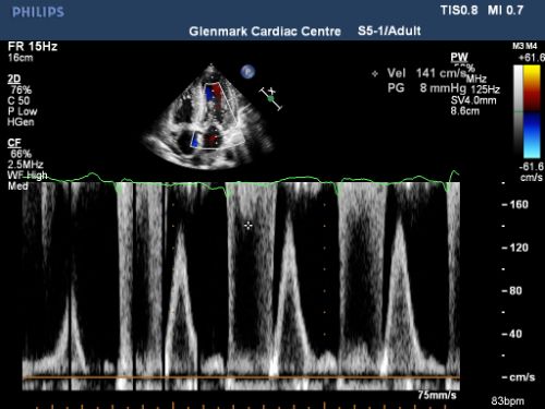
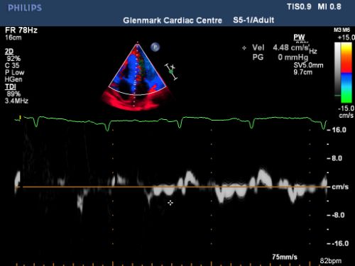
The echo clips and image of PLAX , SAX, apical 4 chamber ,3 chamber and 2 chamber view (Fig A ,B,C, D, E) show a significant LVH which is diffuse and with a speckled appearance. The LV systolic function is normal - LVEF = 0.57 . There is a mild MR and trivial TR. The MV Doppler is suggestive of Type III LV diastolic dysfunction (Fig F, G). There is no regional wall motion abnormality at rest.
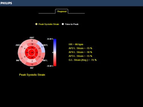
The bulls eye view on Speckle tracking reveals a cherry on the top appearance (Fig H) with a GLS of -15%– suggesting the possibility of Amyloid- restrictive cardiomyopathy
IMP: Infiltrative restrictive cardiomyopathy – Amyloid confirmed on cardiac MRI. Further testing revealed the presence of multiple myeloma
