January 2023
INTERESTING ECHO OF THE MONTH
Case History
A 62 year old male patient came with H/o PTCA and anterior MI in the past, came for routine follow up.
What is the Diagnosis??
The echo clips and image of apical 4 chamber view (Fig A ) There is an echo free space measuring 47 x 16 mm in dimensions seen in the 4 chamber around the apex. There is a regional wall motion abnormality consistent with an anteroseptal MI. In the PLAX view (Fig B ), there appears to be a communication with to and fro flow between this space and the LV at the junction of the anterior septum and the mid septum -SAX view (fig C) suggestive of LV pseudoaneurysm.
IMP : Strong suggestion of large apical LV pseudoaneurysm.
Case History
70 yrs-old, known case of IHD, S/P PTCA & STENT to LAD in 2002 & later CABG (2006). Since then has LV dysfunction (LVEF 0.25). Recently was hospitalized with palpitations & worsening heart failure.
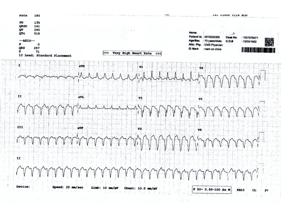
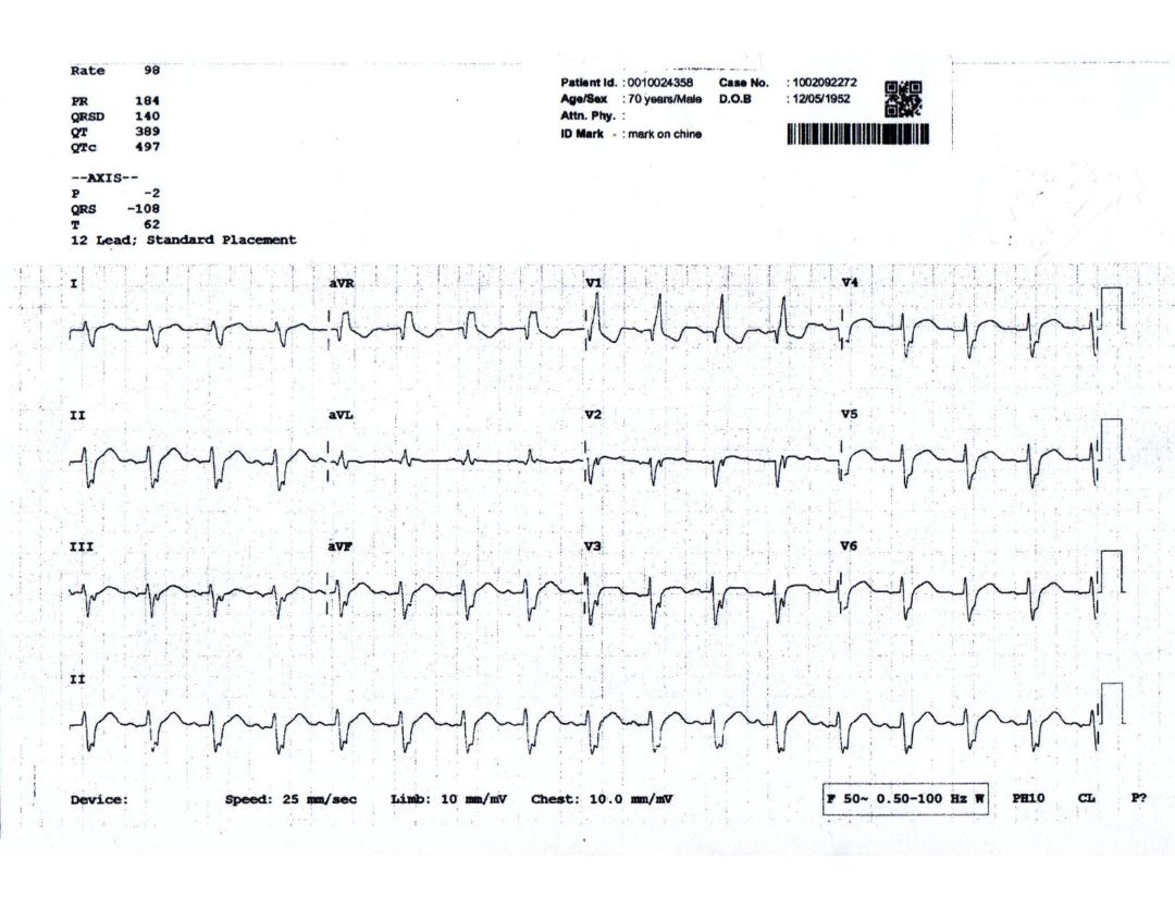
Admission ECG (figure 1) shows wide QRS tachycardia at a rate of 180 bpm. Figure 2, is the sinus rhythm ECG.
Question- Is the wide QRS tachycardia SVT with aberrancy or VT?
The wide QRS tachycardia is VT because:
- More than 90% of all wide QRS tachycardia presenting to the emergency room are VT.
- In a patient with past MI & LV dysfunction, VT is the most likely cause of wide QRS tachycardia. Though the QRS morphology during wide QRS tachycardia is very similar to the SR ECG, suggesting it to be SVT with aberrancy, this is not so because:
- There is AV dissociation. The arrow points to the probable p waves in lead II
- R in avR (in place of qR) and QS in V6 (in place of rS) during wide QRS tachycardia suggests VT


