September 2019
INTERESTING ECHO OF THE MONTH
Case History
A 25 year old male patient was found to be not fit during an application for a job. His echo clips are shown. What is the diagnosis??
Non compaction of LV affecting predominantly the inferior and apical segments with impaired LV systolic function. . The echo clip of the AP 4 Chamber view shows dilated LV with depressed LV systolic contractility. The SAX and apical 2 chamber view highlight the excessive and prominent trabeculations at the LV apex which is so characteristic of the disease.
INTERESTING ECG OF THE MONTH
Case History
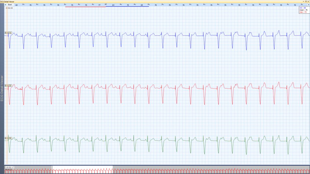
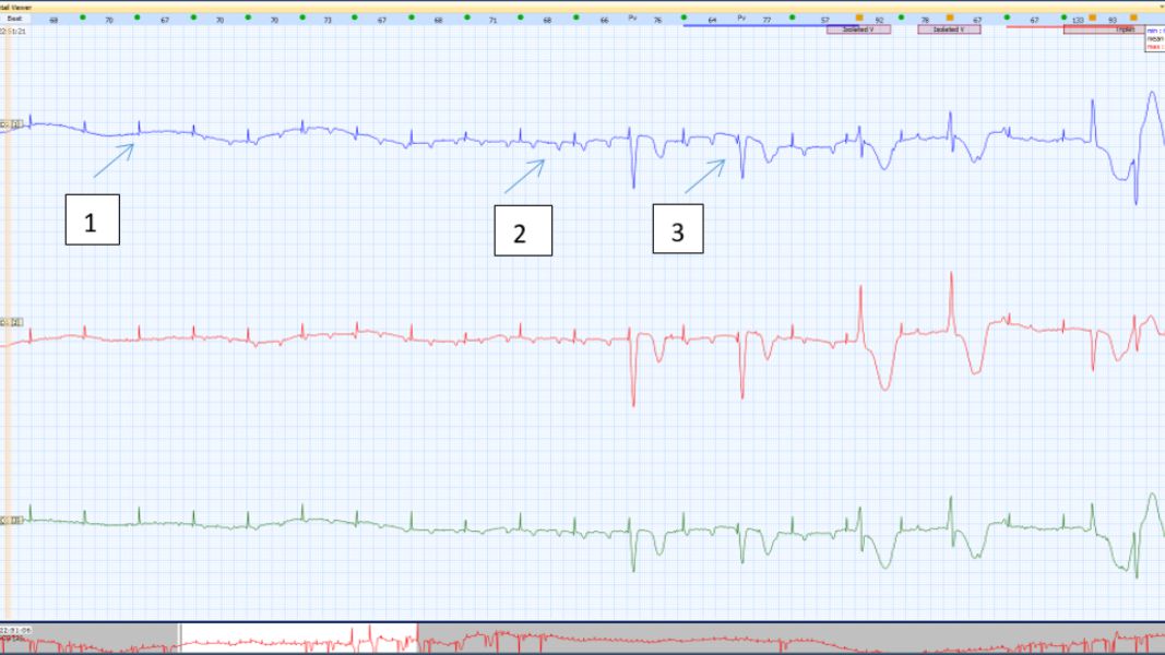
A 86 yrs old male patient with history of pacemaker implantation, was referred for Holter monitoring with history of giddiness. What is the diagnosis?
Pacemaker malfunction with intermittent capture failure. The first Holter tracing shows normal VVI pacemaker function with pacing rate of 70. The second Holter strip shows pacing spike and the heart rate detected by the Holter machine is 72/m, appears normal functioning of the pacemaker. However a close analysis reveals that the pacing spike is not followed by any QRS or T wave and therefore is a clear loss of ventricular capture. There is intermittent loss of ventricular capture. Arrow 1 shows pacing spike not followed by QRS complex, arrow 2 shows atrial depolarization not followed by a QRS complex suggesting high grade AV block. Arrow 3 shows pacing spike followed by QRS complex.


