September 2022
INTERESTING ECHO OF THE MONTH
Case History
A 31 years male was referred for 2 D Echo. He had suffered a myocardial infarct in the past and had undergone PTCA with two stents. Now presented with chest discomfort & dyspnea on exertion. What is the Diagnosis??
Ischemic dilated cardiomyopathy with thrombus in LV and RV
The Echo clips in multiple views show a large mobile thrombus at LV apex & a sessile organized clot in RV apex. There is a regional wall motion abnormality affecting the septum and inferior wall in addition to a diffuse hypokinesia of the other LV segments with severely depressed LV systolic function. EF = 0.20-0.25.
Discussion: Biventricular thrombii are a not a common occurrence. Besides Ischemic heart disease there are other causes of the same. These include biventricular MF,
Loefflers eosinophilic endocarditis, hypercoagulable states such as HIT and APLA, HIV induced cardiomyopathy, Takotsubo cardiomyopathy and other myocarditis.
Treatment will include oral anticoagulation in addition to the other medication
INTERESTING ECG OF THE MONTH
Case History
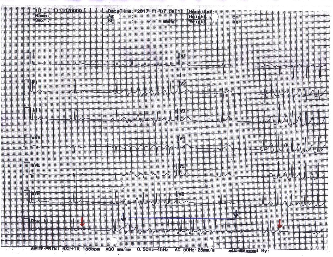
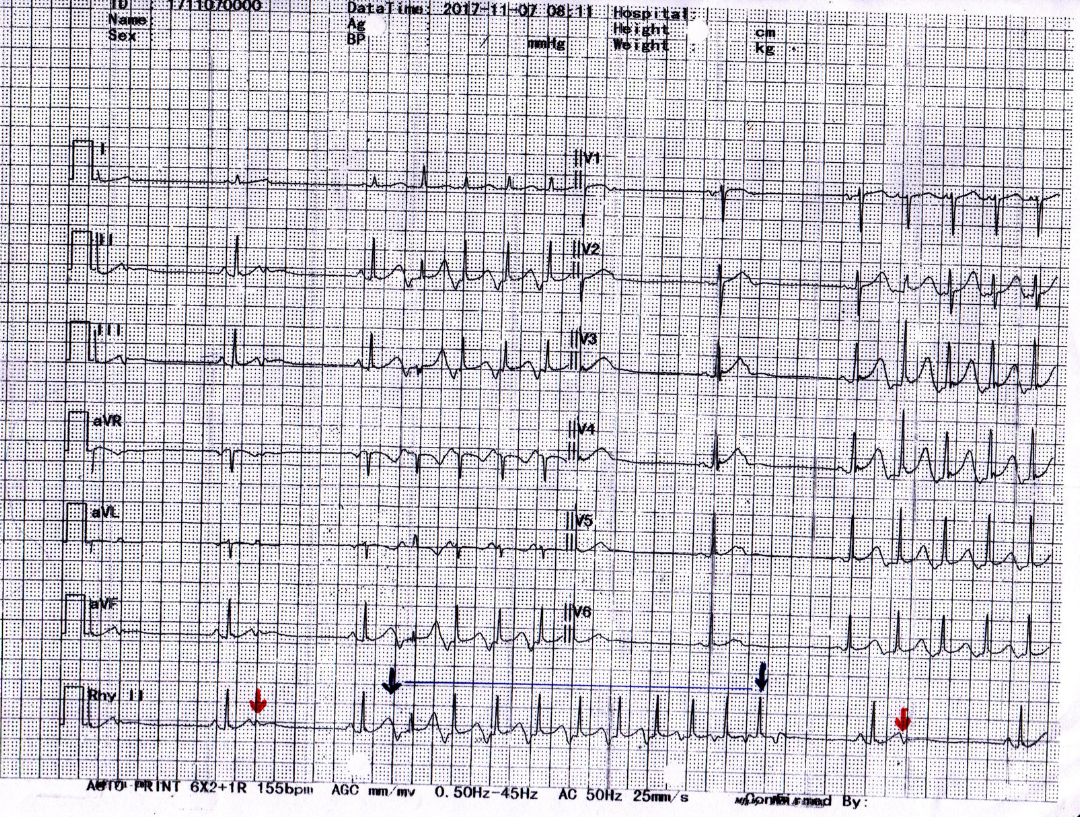
Patient presented with palpitations and giddiness. What does the ECG show?
ECG shows blocked atrial ectopics (red arrows) followed by pauses of around 1.4 seconds along with a run of non-sustained atrial tachycardia (episode between two blue arrows).
The ectopic P wave gets blocked as it falls into the refractory period of earlier beat followed by an apparent pause. One may mistake this for tachy- brady syndrome and hesitate to give antiarrhythmic drugs fearing worsening of bradycardia and pauses. But in this case, the primary problem is atrial ectopics and atrial tachycardia. So this patient must receive antiarrhythmic drugs which will suppress atrial ectopics and thereby take care of atrial ectopics and atrial tachycardia. So, the pauses associated with atrial ectopics will also disappear.


